07. February 2022
AG member Nader Aldoj with a publication on fully automated quantification of prostate zones using magnetic resonance
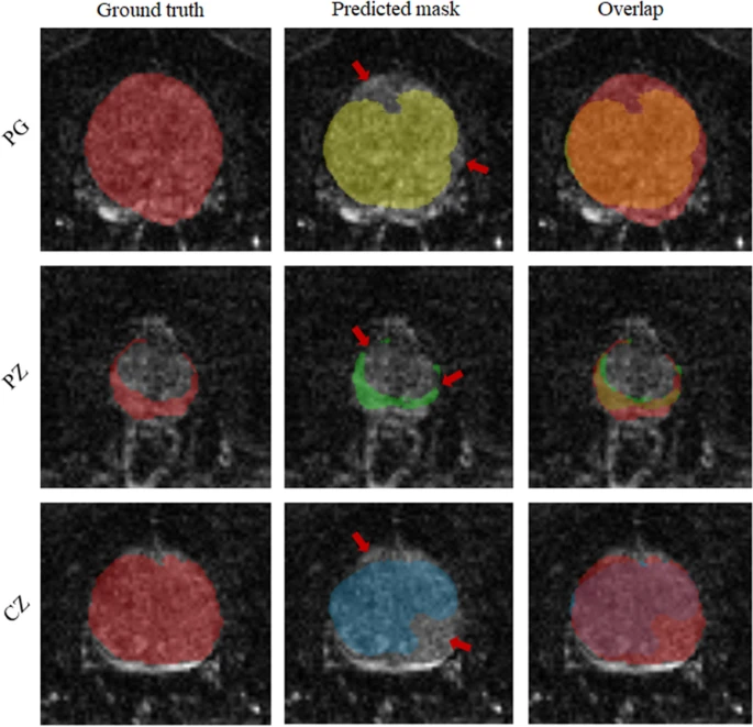
Congratulations to our Team member Nader. Magnetic resonance elastography combined with Dense U-net segmentation allows tabulation of quantitative imaging markers without manual analysis and independent of other MRI sequences and can thus contribute to PCa detection and classification.
Quotes form abstract:
“Using trained network-based image segmentation, we investigated if Magnetic resonance elastography data is suffice to extract anatomical and viscoelastic information for automatic tabulation of zonal mechanical properties of the prostate.” “In conclusion, MRE combined with Dense U-net segmentation allows tabulation of quantitative imaging markers without manual analysis and independent of other MRI sequences and can thus contribute to PCa detection and classification.”
Read the whole article published in nature: https://www.nature.com/articles/s41598-022-05878-5
28. January 2021
Prof. Dewey elected – congratulations!
The results of the 2022 ESR Electronic Elections are in! Congratulations to Andrea Rockall, Christian Loewe, Marion Smits and to our Prof. Marc Dewey on their new positions – Prof. Dewey was elected as the ESR Publications Committee Chair!

Find here the Detailed results of the ESR Electronic Elections 2022.
13. December 2021
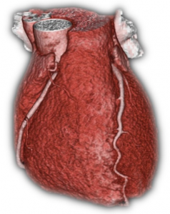
AG member Philipp Karius finished dissertation
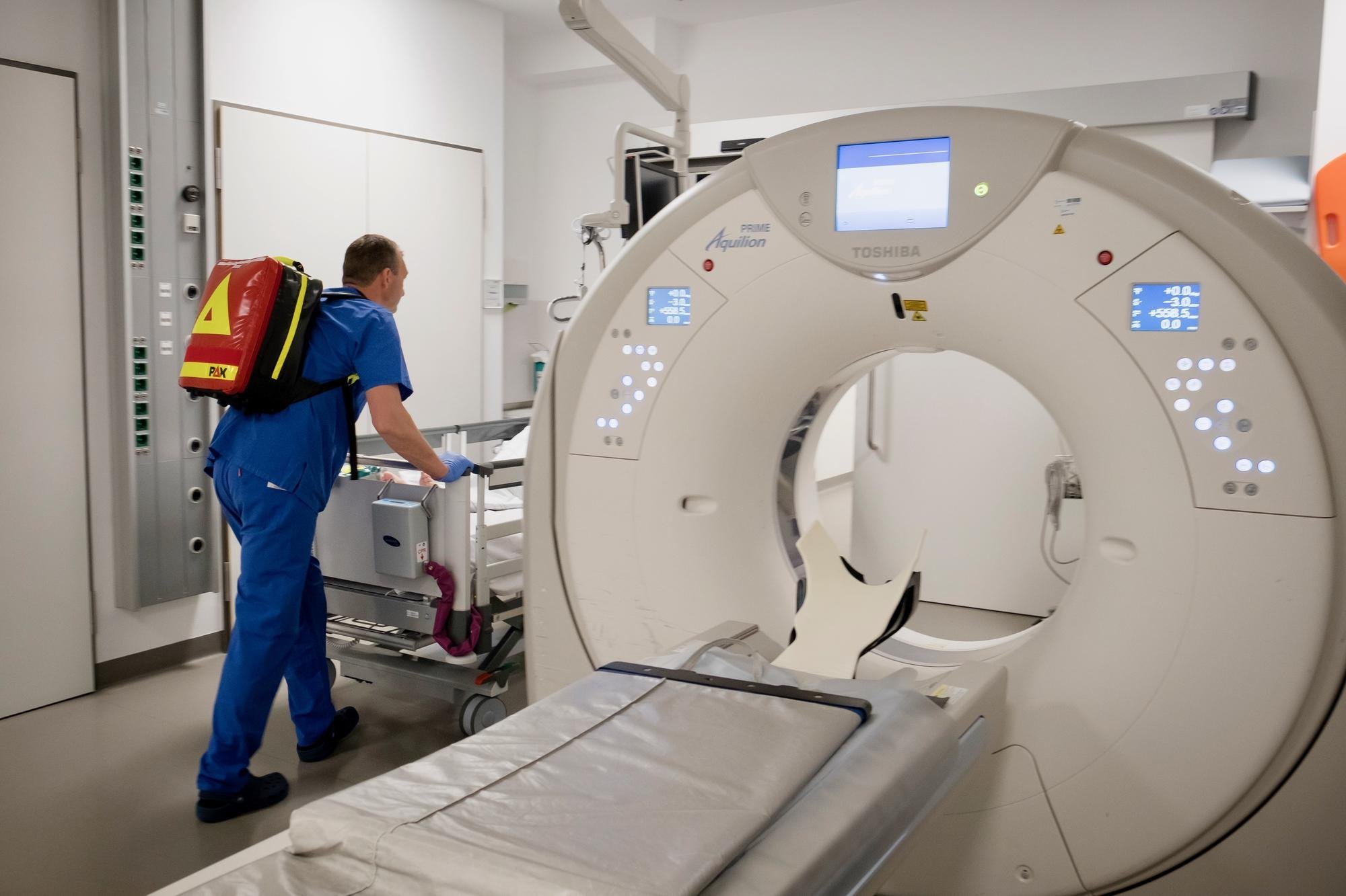
Philipp Karius, a member of our working group, has finished his dissertation. He examined in detail the relevance of extracardiac findings on Coronary CT. Its structured analysis is beneficial in many instances. On the one hand it offers the possibility to detect acute extracardiac pathologies as well as early-stage cancer. On the other hand it can help to find the cause of chest pain when a significant Coronary Artery Disease is ruled out. Hence, paying attention to extracardiac findings on coronary CT can contribute to more efficiency in diagnosis and treatment. The research resulted in three scientific papers: one was published in the Journal of Cardiovascular Computed Tomography (JCCT) and another two papers were published in European Radiology (EUR).
The thesis was awarded with “magna cum laude”. Philipp Karius would like to deeply thank his supervisor Prof. Dr. Marc Dewey as well as Felix C. Sokolowski and all members of the working group for the inspiration and the exciting time. Meanwhile Philipp Karius works in the Department of Radiology of the Städtische Klinikum Dessau.
To find out more please follow the links below:
https://doi.org/10.1016/j.jcct.2014.04.002
https://doi.org/10.1007/s00330-018-5688-4
https://doi.org/10.1007/s00330-018-5432-0
22. October 2021
First MD/PhD Dissertation
We are pleased to announce the first successful completion of an MD/PhD dissertation in our working group by our long-standing member Benjamin Kendziora. The dissertation deals with computed tomography for the diagnosis of coronary artery disease and magnetic resonance imaging for the quantification of saved myocardium after myocardial infarction. Four scientific papers were published in the British Medical Journal (BMJ), Radiology, the British Medical Journal Open (BMJ Open) and PLOS ONE as part of the dissertation. Additionally, Benjamin Kendziora presented research results on the conference of the Radiological Society of North America (RSNA) in Chicago and the European Congress of Radiology (ECR) in Vienna. In summary, the papers’ results suggest that CT angiography supplemented by CT perfusion can reliably and safely detect coronary stenoses and assess their functional relevance in patients with low to intermediate probability of coronary artery disease. It was also shown that magnetic resonance imaging allows the visualization of salvaged myocardium, allowing prognostic predictions to be made.
The thesis was awarded the highest grade “summa cum laude.” Benjamin Kendziora would like to thank his first supervisor Prof. Marc Dewey and his second supervisors Dr. Matthias Rief and Prof. Peter Schlattmann as well as the whole team of the research group for the educational and enjoyable time. In the meantime, Benjamin Kendziora is working at the Department of Dermatology at the Ludwig Maximilian University of Munich as a resident physician in further training. We will continue to work together and look forward to upcoming scientific projects!
Please find more information on the publications below:
http://doi.org/10.1136/bmj.i5441
http://doi.org/10.1148/radiol.2017162447
http://doi.org/10.1136/bmjopen-2019-034359
http://doi.org/10.1371/journal.pone.0228736
13. September 2021
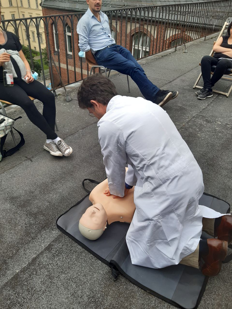
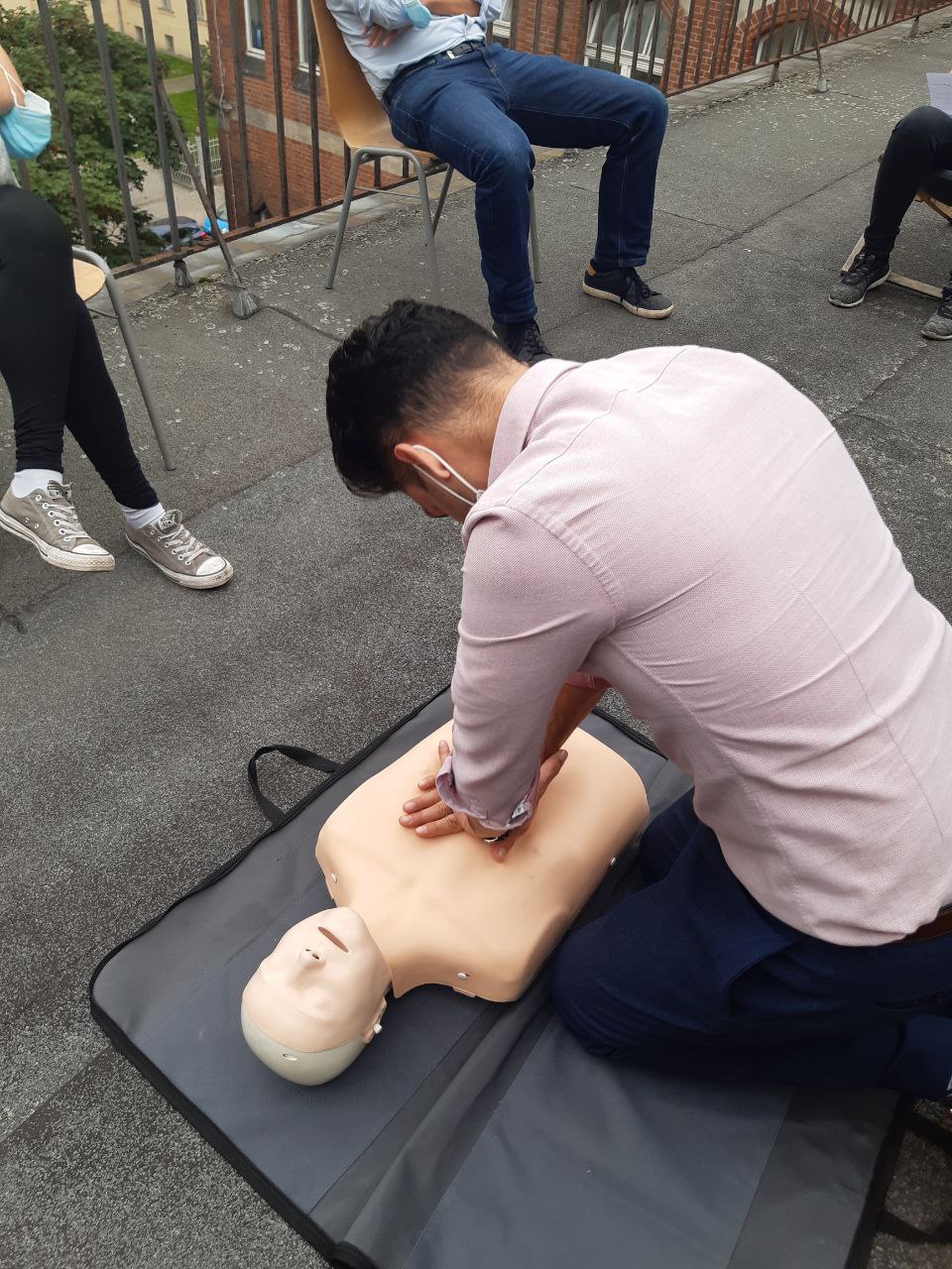
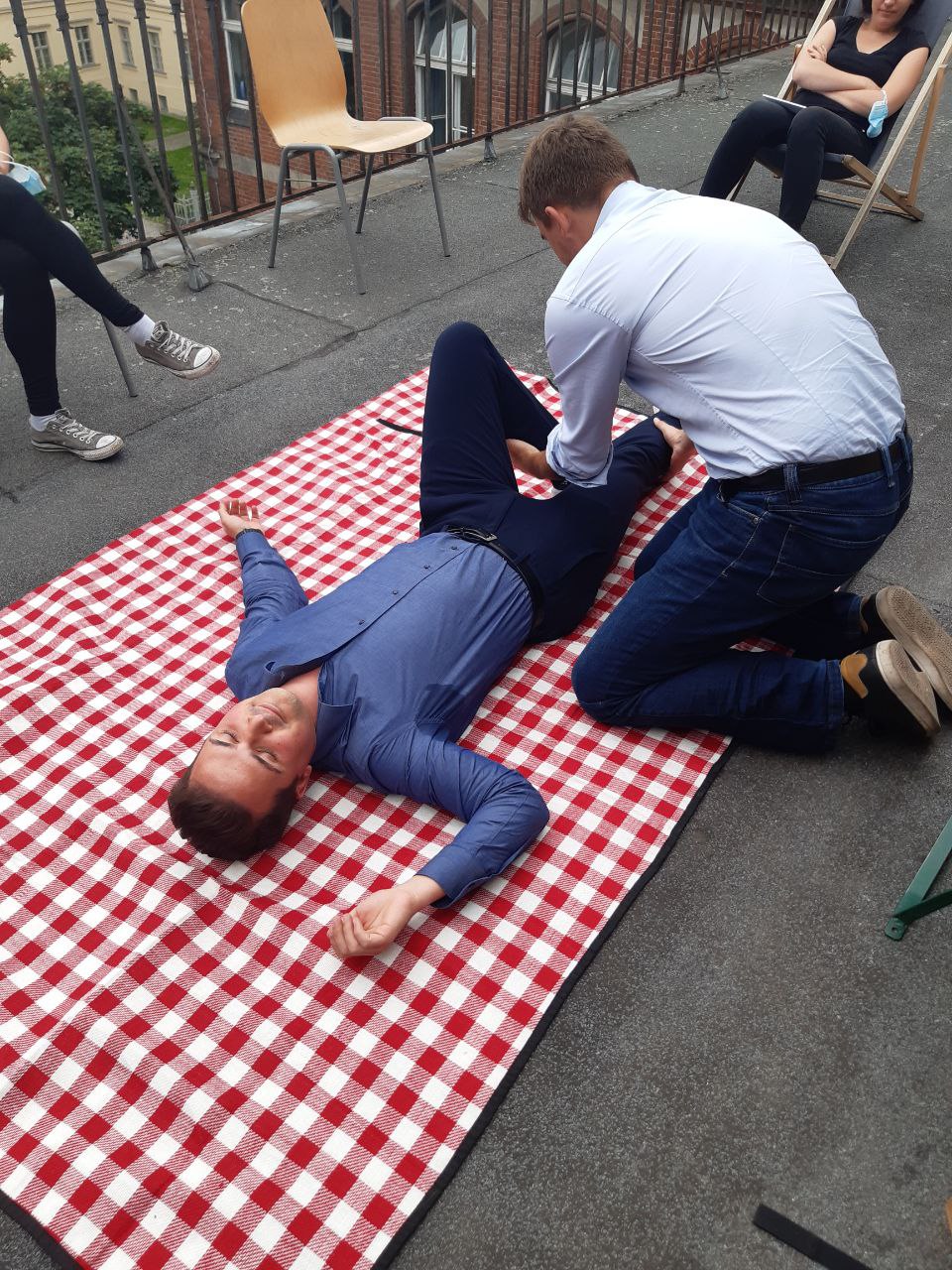
First Aid Course by Promotionskolleg member Alina
Today it was the time to freshen up our rescuing skills. Alina, which happens to be a tutor of the Charité Promotionskolleg and also a first aid course instructor, reminded us on how to react properly in emergency situations, where every second counts!
Resuscitation was of course on the menu (you remember: 5cm + 30:2 + “Staying alive” 😉 as well as bandaging. Lots of fun and feeling ready for a situation like this! Thank you very much Alina!
12. July 2021
New publication on fully automated quantification of prostate elasticity
Our @team_dewey group member Nader Aldoj just submitted his work to #arxiv by the Cornell University. This research has been done with the help of our colleagues Federico, Anja, Patrick and Ingolf and funded by @dfg_public.
Abstract:
Magnetic resonance elastography (MRE) for measuring viscoelasticity heavily depends on proper tissue segmentation, especially in heterogeneous organs such as the prostate. Using trained network-based image segmentation, we investigated if MRE data suffice to extract anatomical and viscoelastic information for automatic tabulation of zonal mechanical properties of the prostate. Overall, 40 patients with benign prostatic hyperplasia (BPH) or prostate cancer (PCa) were examined with three magnetic resonance imaging (MRI) sequences: T2-weighted MRI (T2w), diffusion-weighted imaging (DWI), and MRE-based tomoelastography yielding six independent sets of imaging data per patient (T2w, DWI, apparent diffusion coefficient (ADC), MRE magnitude, shear wave speed, and loss angle maps).
06. July 2021
Interested in a doctoral thesis on medical imaging using #fractalanalysis?
Announcement Student Assistant 40h/month
We are looking for energetic support for our team in the EU-funded pan-European and multicenter DISCHARGE study. It is a pragmatic randomized controlled trial comparing effectiveness of cardiac CT with cardiac catheterization in patients with stable chest pain and suspected coronary artery disease (CAD). The study will include a cost analysis for which we are seeking medical students with an interest in this topic area as well as a PhD.
We are offering a position as a student assistant based on 40 hours per month for a minimum of 6 months starting immediately. Our office is located at the central campus. We are looking for motivated students who are interested in clinical trials, enjoy working in a team and are willing to take on responsible tasks and ideally have some prior experience in medical cost reporting.
Your tasks:
- Support of the cost team
- Literature research
- Data preparation
- Communication with participating study centers and responsible project partners.
We are looking for:
- Medical students with interest in pursuing a PhD
- Ideally previous experience in the field of medical cost recording
We offer:
- As a coordinating center of the EU-funded multicenter DISCHARGE study, an interdisciplinary team at the Campus Mitte.
- Remuneration according to the collective agreement for student assistants 40h/month for 6 months
- Working hours can be arranged flexibly according to our core working hours.
If you are interested or have any questions, please send your application by mail to: discharge.eu@charite.de
Further information about the study: https://www.dischargetrial.eu/de/
15. June 2021
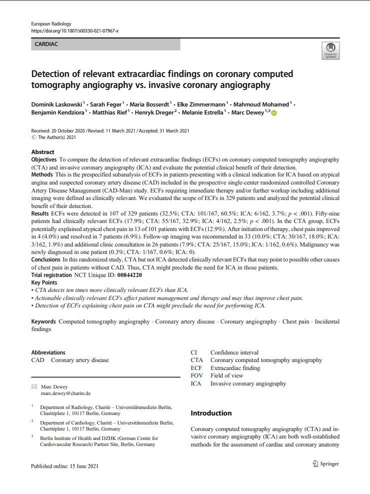
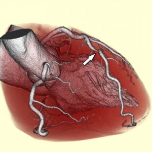
New publication: Subanalysis on CTA vs. ICA on patients with atypical angina
Detection of relevant extracardiac findings on coronary computed tomography angiography vs. invasive coronary angiography.
While both coronary computed tomography angiography (CTA) and invasive coronary angiography (ICA) are well-established methods for the assessment of cardiac and coronary anatomy, they visualize also surrounding structures, such as lungs, mediastinum, and upper abdomen, leading to detection of extracardiac findings (ECFs). We performed a subanalysis of clinically relevant ECFs on CTA versus ICA in patients with atypical angina and suspected coronary artery disease (CAD) based on data from our randomized controlled CAD-Man trial. Our study showed that CTA detects ten times more clinically relevant ECFs than ICA. Some of those ECFs detected by CTA (13%) were identified as explaining patients’ chest pain and may thus affect management and therapy and consequently improve quality of life. Detection of ECFs explaining chest pain on CTA might also preclude the need for performing ICA, which is invasive and associated with more procedural complications than CTA.
Read more: https://link.springer.com/content/pdf/10.1007/s00330-021-07967-x.pdf
12. June 2021
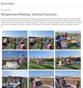
Finally there: all pictures from our perspectives meeting / team building event
See more: marcdewey.de/excursions
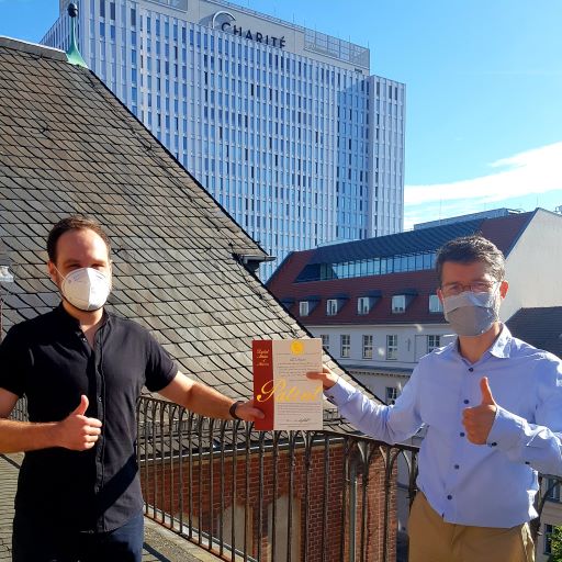
10. June 2021
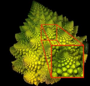
Method for characterizing perfusion abnormalities by means of fractal analysis of the interface region
Dr. Florian Michallek just released details of our patent on #fractal analysis of perfusion imaging based on self-similarities often found in nature. A great example is the romanesco broccoli also exhibiting scale-invariant self-similarity. We apply fractal analysis to #CT and #MRI.
09. June 2021
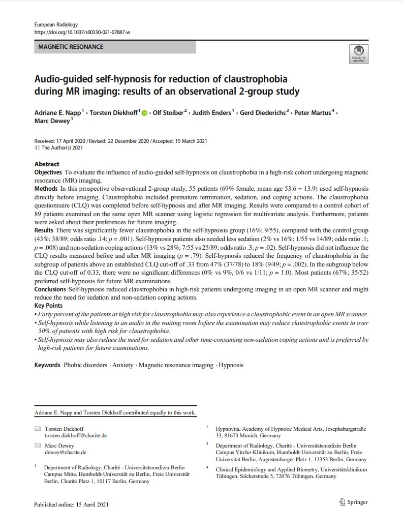
Collaboarative Article released on “Audio-guided self-hypnosis for reduction of claustrophobia during MR imaging”
Great work by Dr. Adriane Napp and Torsten Diekhoff from our Department of Radiology as equal first authors in collaboration with amazing Olf Stoiber from Hypnovita (München). Special thanks also go to Judith Enders, Gerd Diederichs (Department of Radiology) and Peter Martus (Universitätsklinikum Tübingen). Their article “Audio-guided self-hypnosis for reduction of claustrophobia during MR imaging” has been published successfully on April 17th, 2021 by European Radiology. A summarizing podcast has been written by Torsten Diekhoff and an editorial about the topic in European Radiology.
About 10% of the patients undergoing MRI experience a claustrophobic event which is defined by the need for either sedation or other coping actions to complete the examination. Incomplete examinations due to claustrophobia are also regarded as an event. The use of audio-guided self-hypnosis enabled the reduction of events by half. Thus, this approach is very promising to follow-up on for managing claustrophobia during MRI.
The article can be found under: Article (springer.com)
The editorial can be found under: Editorial Springer Link
The podcast can be found under: Podcast/ Twitter

05. June 2021
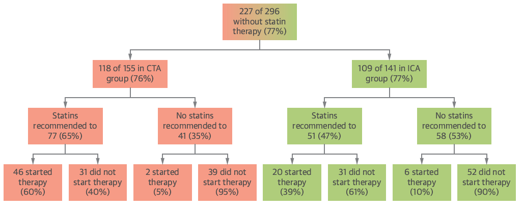
Effects of cardiac CT on statin intake and cholesterol levels
Sub- analysis of the randomized controlled CAD-Man trial on the effect of coronary CT angiography on statin adherence published by Sarah Feger and Laura Elzenbeck in JACC Cardiovascular Imaging.
The study showed that an intervention based on computed tomography versus invasive coronary angiography resulted in improved statin adherence and cholesterol levels.
After a median follow-up of 3.3 years, more patients in the CTA group compared with the ICA group (60% versus 39%) reported to be on statin therapy. Total cholesterol and LDL cholesterol levels were decreased in the CT group (decrease by 22.6% and 35.5%), but increased in the ICA group (by 19.8% and 28.9%).
01. June 2021
Prof. Dewey interviewed about confidant role
Not only to ensure the high quality of dissertations or to prevent doctoral students from dropping out, the Charité has created several offers to support doctoral students and supervisors. These also include the activities of the confidant in a short interview, Professor Dr. Marc Dewey, deputy director of the Institute of Radiology at the Charité Campus Mitte, tells us what their tasks as confidants include (Article in German language)
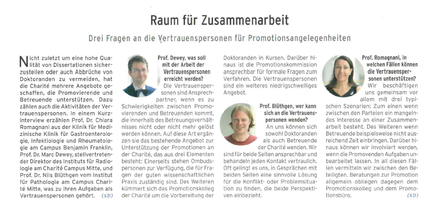 (Click to enlarge article)
Thank you for the interview.
19. May 2021
Prof. Dewey interviewed by Zeit on Hypnosis vs. Anesthesia

“Germany’s best-known university hospital is experimenting with hypnosis techniques. In the ZEIT WISSEN podcast, the doctors report on their initial findings.”
Prof. Dewey in the “Zeit Wissen” Podcast about research on Hypnosis as a Anesthesia alternative.
Read & listen more:
Hypnose: Hypnose statt Narkose – funktioniert das? | ZEIT ONLINE
28. January 2021
AuntMinnie nominates Prof. Dewey as a finalist!
Congratualations to Prof. Dewey as being chosen by the number one medical imaging community as a finalist for the most influential radiologist! Fully deserved!
07. January 2021
A New Family Member!
The entire workinggroup sends our big congratulations to our dear member Heli on her 4th kid! We find it Wonderful and hope to see it live, as soon as the situation allows. Of course we hope the new AG-baby will step into its mothers footsteps and become a great medical doctor or scientist as her mother!
Prof. Dewey wishes Heli all the best and is also looking forward to see the our new member on our weekly online meetings.
See you soon!
17. December 2020
Thank you for this lovely gift from one of our students
A great gift from our student Helene Wunderlich who put time and effort into this little CT model as a little thank you present to Prof. Dewey and Viktoria Wieske who supervised Helene during her time in our group.
Thank you for this lovely and creative present. And thank you for your work in our group and inside the Discharge-Team!
10. December 2020
New European Journal of Radiology publication: Can CT help in patients with obscure infections?
Our working group Member Dr. Julian Pohlan just published a research in the European Journal of Radiology on predicting possible sources of infections.
Results:
In 133 out of 196 (67.9 %) body CTs from general wards with severe infection or sepsis, body CT identified an infectious focus. 90 % of the infections were located in the chest, abdomen, and genitourinary tract, in descending order. In 76.5 % (150 of 196) of examinations, CT correctly predicted the final infectious source. The positive predictive value (PPV) of a CT-detected focus was 84.2 % (95 % CI 79.0%-88.3%).
Read more about this research in the European Journal of Radiology.
Congratulations to Dr. Pohlan!
04. September 2020
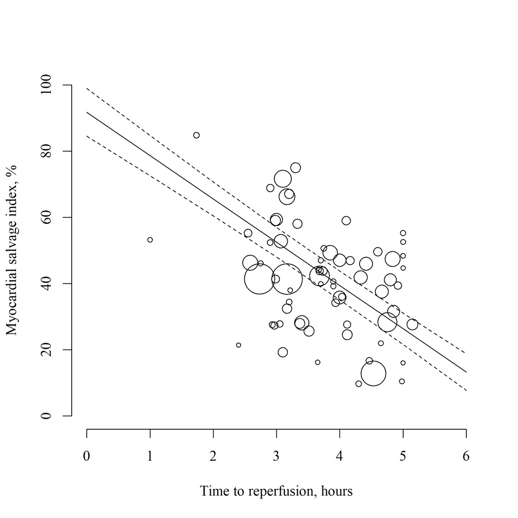
MRI for measuring therapy efficiency after revascularisation in ST-segment elevation myocardial infarction
In this systematic review and meta-analysis, Benjamin Kendziora et al. analysed whether myocardial resonance imaging can be used to measure therapeutic efficiency after myocardial infarction. Indeed, the data suggest that the myocardial salvage index calculated by quantifying post-infarctional myocardial oedema and necrosis with myocardial resonance imaging provides information on therapeutic efficiency.
Future research in the field will be interesting, particularly with regard to mapping of the heart by myocardial resonance imaging, which increases comparability across hardware and software systems.
The work is freely available here:
https://bmjopen.bmj.com/content/10/9/e034359

29. September 2020
A collaborative meta-analysis of the clinical usefulness of coronary CT angiography
Viktoria Wieske and Marc Dewey jointly provided a review article for the summer issue of the Diagnostic Imaging Europe Magazine about our worldwide COME-CCT Consortium and the primary results published in BMJ last year lead by our working group colleague Robert Haase. Following our main analysis publication and especially in regards to the new European Guideline on chronic coronary syndrome we are currently very much looking forward to more interesting and exciting publications from our COME-CCT project to come soon. Stay tuned!
The review article in Diagnostic Imaging Europe can be found here:
https://www.dieurope.com/site/wp-content/uploads/2020/07/DIEurope-JuneJuly-2020.pdf
15. September 2020
Insights into clinical pre-test probability for CAD
Our working group member Dr. Sarah Feger just released the results of her work – we’d like to congratulate on that!
The objective of this paper was nothing less, than to test the accuracy of clinical pre-test probability (PTP) for prediction of obstructive coronary artery disease (CAD) in a pan-European setting. In total, 1440 patients (654 female, 786 male) were included at 25 clinical sites from May 2014 until July 2017.
Outcome:
• Clinical pre-test probability calculation using the initial and updated D+F model overestimates the prevalence of obstructive CAD identified by ICA and CT.
• Overestimation of disease prevalence is higher for the initial D+F compared with the updated D+F.
• Diagnostic accuracy of PTP assessment varies strongly between different clinical sites throughout Europe.
Read the full paper here.
04. September 2020
Patient preferences for development in MRI scanner design: a survey of claustrophobic patients in a randomized study
Claustrophobia is a common problem in Magnetic Resonance Imaging (MRI)-examinations: up to 10% of patients suffer from it. Finding satisfying solutions for prevention and management of claustrophia in MRI-exams remains a challenge.
So what do claustrophobic patients themselves suggest to improve their MRI-experience? We asked claustrophobic patients as part of a randmozied controlled trial what MRI-scanner design they prefer.
Our results:
Patients at high risk to experience claustrophobia in the MRI want open as opposed to closed scanner bore-designs.
They want less noise during the examination.
And interestingly, they show an increased acceptance of closed-bore MRI-designs, if they are specifically educated about the higher diagnostic performance of this design.
This information can be useful for referral of claustrophobic patients as well as future scanner development.
01. September 2020
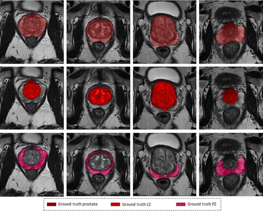
Automatic prostate segmentation
A new step toward an improved automatic prostate and zones segmentation. This piece of work discusses a new neural network architecture (Dense 2 U-net) based on the state-of-the-art U-net, and highlights its pros, cons and added values.
We trained the algorithm on 141 patient datasets and tested it on 47 patient datasets using axial T2-weighted images in a four-fold cross-validation fashion. The networks were trained and tested on weakly and accurately annotated masks separately to test the hypothesis that the network can learn even when the labels are not accurate.
Read more: https://www.nature.com/articles/s41598-020-71080-0
21. August 2020
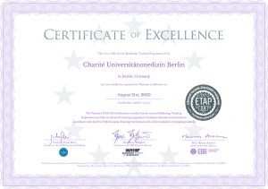
Department of Radiology at Charité Awarded Platinum
On August 21st 2020, the Department of Radiology at Charité became the first German department to have been awarded a platinum certificate of the European Training Assessment Program (ETAP) of the European Board of Radiology (EBR). This is a Europe-wide standardized certification which has been accredited to our radiology training program.
Departments accredited with the platinum award – the highest possible attainable certificate – are for those aligned to the European Society of Radiology (ESR) European Training Curriculum, characterized by the availability of specialized advanced training staff, excellent and personal supervision of advanced training residents, a mentoring program, excellent training in emergency radiology, the involvement of residents in interdisciplinary team meetings, and the opportunity to work in science, management, and leadership roles, among other criteria.
27. June 2020
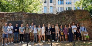
Summer excursion: Nikolaiviertel + coffee tasting
On a very summery day in June, the Dewey workgroup started an excursion to the beginnings of the city of Berlin. But not without first having an energizing espresso tasting. Wild Espresso Coffee from Punjab served as the starting signal for this excursion. Rolf Gänsrich, journalist, freelance author and connoisseur of Berlin’s history, guided us through the Nikolai Quarter, which belongs to the oldest core of the city and under which the roots of Berlin’s settlement lie. The tour ended with a visit to the old city wall of Berlin, whose remains from the 13th century are still partly preserved, and the imposing ruins of the Franciscan monastery church.
Thanks to all participants for a fun and interesting time, we are looking forward to next year!
[ngg src=”galleries” ids=”6″ display=”basic_imagebrowser”]
June 11, 2020
Congratulations to Mr. Federico Biavati!
Federico Biavati has successfully passed the state examination. Congratulations! He will now work on his MD/PhD with Bernhard Föllmer in BIOQIC
June 09, 2020
Answers to artificial intelligence in radiology
Artificial intelligence in radiology: what do patients, doctors, radiographers and AI developers say? The answers are finally here from our @BerlinUAlliance Project with @gersch_martin, Birgit Beck from @TUBerlin, Jenny Wesche form @FU_Berlin and Annekatrin Hoppe from @HumboldtUni.
Read more: berlin-university-alliance.de/en/impressions
June 05, 2020
Prostate cancer detection by deep learning?
Does deep learning work for prostate cancer detection on MRI? It appears yes, a multi-channel 3D CNN was as accurate as reported for radiologists. Full paper published in print by Nader Aldoj from our team.
Read more: link.springer.com/article/10.100
June 05, 2020
Noise Reduction through Deep Learning
Reduction of noise or artifacts is very important in medical image reconstruction. Using a novel generalized deep learning-based approach our Alumni Andreas Kofler from the BIOQIC program, now at PTB, could improve CT and MRI.
Read more: iopscience.iop.org/article/10.108
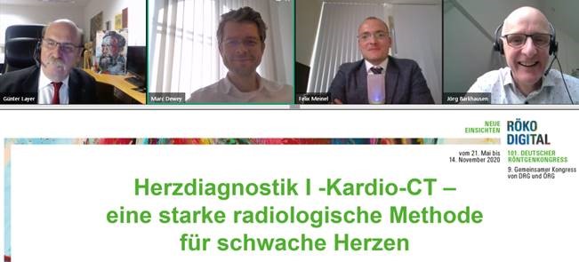
May 21, 2020
Röntgenkongress online
Holiday, 25 degrees, sun and: 350 spectators at the online Röntgenkongress today about heart imaging. Thanks to @DRG_en for the organisation and to Jörg Barkhausen, Günter Layer and Felix Meinel for the good atmosphere!
May 19, 2020
Findings about life quality of women with coronary artery disease
The following results were just released by PD Nina Rieckmann and Prof. Dewey after analysing the DISCHARGE pilot study data. Women have worse quality of life compared with men if they are suspected of having coronary artery disease.
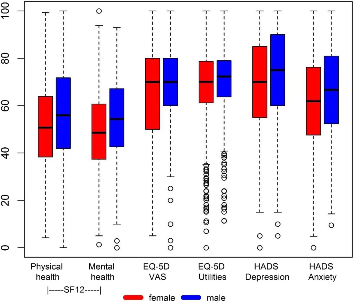
Read about the findings ont the BMC page here.

May 4, 2020
Charité named #5 in top hospital list
Congratulations to the Charité Universitätsmedizin Berlin!
Ranked number 5 in Newsweek’s list of best medical centers is a thing everybody can be proud of. Plus the fact that Charité leads by far with their amount of 3011 hospital beds, makes this achievement even more impressive! Also as Newsweeks editor in Chief Nancy Cooper remarked: “…the researchers and experts who help build this authoritative list are keenly aware of what’s going on in hospitals around the globe, and they can, with confidence, identify the ones that set the standard for excellent care…” we’d like to congratulate every hospital on the list for providing such an excellent quality of medical care. Team Dewey is proud of being part of this great organisation and of being able to contribute to this result.
Link: www.newsweek.com/best-hospitals-2020

April 22, 2020
How to get rid of artifacts in MRI
Convolutional neural networks using our XT,YT-approach developed by our working group member Andreas Kofler are a viable option to have cardiac cine MRI without artefacts even with small training datasets. Our method outperforms the 2D spatially trained U-net and the 2D spatio-temporal U-net. Compared to the 3D spatio-temporal U-net, our method delivers comparable results, but requiring shorter training times and less training data.
April 08, 2020
Congratulations Adriane Napp
Our dear working group Member Adriane E. Napp released her work in European Radiology and we’d like to congratulate her on that. Together with Ginaluca de Rubeis she researched on image quality and protocol adherence of computed tomography and invasive coronary angiography. See the full article here.
March 20, 2020
Covid 19 Info links
Dear readers, in order to help stopping the pandemic, we have set up an informational page with resoruces regarding Covid-19. There is a lot of information circulating, these links are from sources we trust. It is important to us to help ensure that the right information reaches people.
See all information on Covid 19 here.
February 25., 2020
Myocardial Ischaemia Consensus published!
Great news! After a big load of work and time, finally the “Myocardial Ischaemia Consensus” was published in Nature Reviews Cardiology.
Check it out here: https://www.nature.com/articles/s41569-020-0341-8
February 12, 2020
A friendly new face for our tea
We’d like to give a heartly and warm welcome to a new member of our team: Eser Isler is a physician and docotral student hailing from turkey. She is not only extraordinarily interested in medicine, but also in yoga and python – and since that is not enough, she made it on the 16th place in the “Higher Education Entrance Exam (YGS-2012)” – which almost 2 Million students attended, Graduated from Hacettepe University Faculty of Medicine. Congratulations on your efforts and welcome to AG Dewey!
January 27, 2020
January 8 & 9, 2020
Social Cohesion and Medical Imaging Meeting
The kick off meeting for the work on Social Cohesion and Medical Imaging within the “Berlin University Alliance”, in which Prof. Dewey is the main applicant, took place on 8. & 9. of January in the “Window of Science” in the CCO at the Charité Campus Mitte. We are looking forward to the results of this promising cooperative project.
January 7, 2020
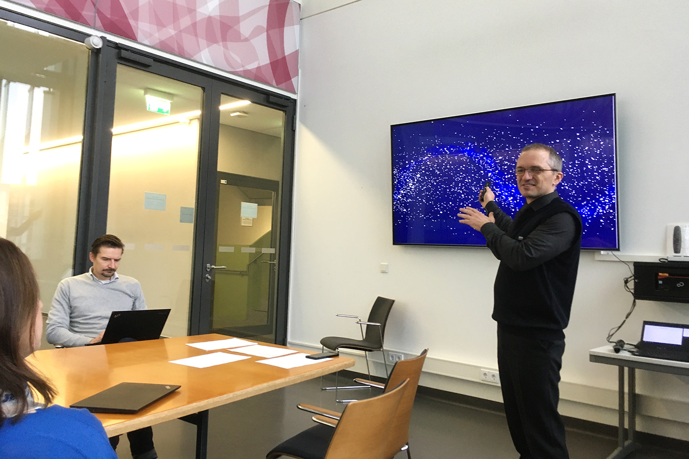
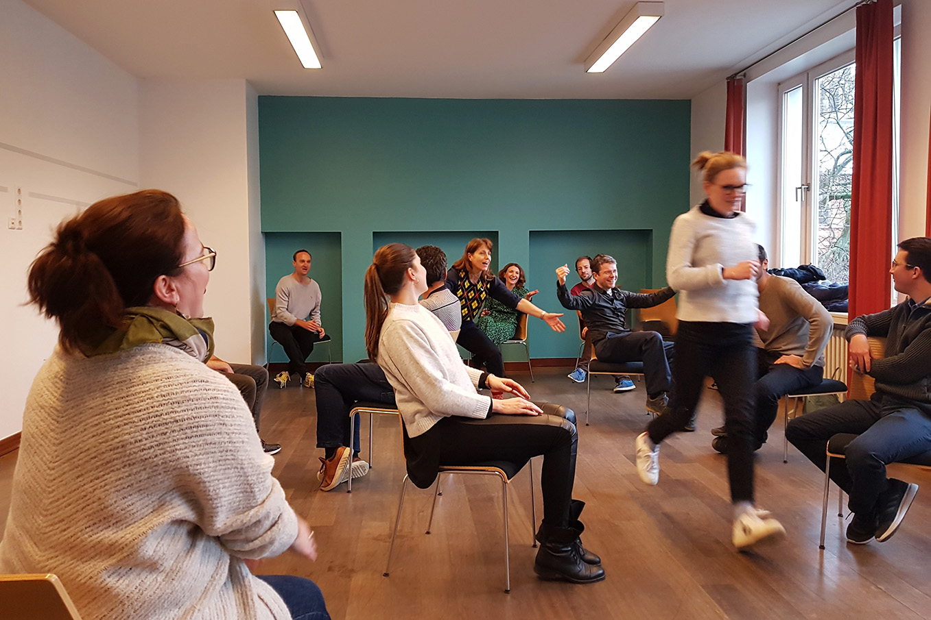
Groups Dewey on perspective meeting/Teambuilding
In order to start strong into the new year, the group of Prof. Dewey conducted a perspective meeting including a fun and mind opening teambuilding event. Not only the whole groups mental skills were asked, but also the physical ones (as you can see in the picture) from time to time at this whole day event. We thank for the invitation and the great idea and look forward to take on every challenge of the new year!

December 4, 2019
Prof. Dewey now on twitter
If you’re on Twitter, the fast social media platform for news and communication, make sure to check out and follow Prof. Dewey: @ProfDewey.
He is tweeting about #medical #imaging #radiology and everything related and looking forward for an exchange via Twitter with you.
November 12, 2019
Results on clinical trials in radiology and data sharing
In this work of Dr. Maria Bosserdt, Prof. Bernd Hamm and Prof. Marc Dewey randomised trials were analysed, since they are a key contributor to evidence-based medicine and radiology. They play an increasingly important role in developing evidence-based strategies for diagnostic management and image-guided treatments.
Scientific discoveries will increasingly depend on sharing data on a global scale. While data sharing is rather common in life sciences, data sharing is lagging behind in clinical medicine including medical imaging. Medical imaging and radiology play a central role in patient care and typically create very large data sets, which are challenging to curate, share, and reanalyse . Thus, we conducted an online survey among European heads of imaging departments and speakers at the Clinical Trials in Radiology sessions at the European Congress of Radiology (ECR) 2015-2018 to ascertain the current situation and anticipate future directions of imaging trials and data sharing in radiology.
Read all about the results here.
November 12, 2019
Prof. Dewey in correspondence about artificial intelligence in The Lancet
Prof. Marc Dewey and Peter Schlattmann went into correspondece with The Lancet, their letter regarding the study on the diagnostic application of deep learning algorithms in head CT imaging has been answered by The Lancet already. Read the reply on the warning about possible obstacles in clinical workflows caused by the use of artificial intelligence here.
The current editorial of The Lancet can be found here.
November 09, 2019
Artificial intelligence in radiology
Today, the first meeting of the Artficial Intelligence in Radiology working group took place. This is in preparation of the first Research Forum of the Berlin University Alliance.
November 06, 2019
Welcome Zuse Institute
We had a visit of the Zuse Institute of Berlin under lead of Prof Hege at our research group on October 29th. Great sharing of experiences and ideas. We are looking forward on cooperating together in the future!
The Zuse Institute Berlin (ZIB):
is an interdisciplinary research institute for applied mathematics and data-intensive high-performance computing. Its research focuses on modeling, simulation and optimization with scientific cooperation partners from academia and industry.
September 23, 2019
Successful PhD thesis defense on Astra4D
Today, Steffen Lukas (pictured on the left together with his supervisor Prof. Dewey) successfully defended his PhD thesis on ASTRA4D motion registration with the highest score of summa cum laude. Congratulations!
September 13, 2019
Funding in the DFG priority programme Radiomics
Over the next three years, the DFG will provide eight million euros to fund a priority programme on radiomics with a total of 17 projects.
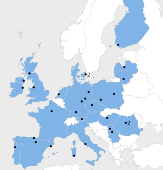
The working group Prof. Dewey has successfully acquired two of these projects and will carry out new analyses of coronary plaques and coronary flow with data from over 3500 patients from the DISCHARGE study (www.dischargetrial.eu).
September 12, 2019
Feature in Radiology Podcast
The most recent publication by our dear AG members Maria Bosserdt, Elke Zimmermann and others entitled “Kidney Injury after Intravenous versus Intra-arterial Contrast Agent in Patients Suspected of Having Coronary Artery Diseas” has found its way into the RADIOLOGY podcast.
The podcast: Feel free to listen here (From minute 17:11 on).
The transcript: can be found here (At page 3).
More Radiology podcasts can be found here.
We would like to thank the editorial board of RADIOLOGY for featuring our work!
September 6, 2019
Charité Cardiac CT Course
Even Aunt Minnie recommends it! The Charité Universitätsmedizin Berlin radiology department is hosting a hands-on cardiac CT course (www.ct-kurs.de) from 16. to 20. September, and a limited number of spaces are still available.
About the course:
There have been dramatic technical advances in computed tomography for imaging of the heart and particularly the coronary arteries. During the course you will had 100 hands-on cardiac CT cases with invasive angiography correlation. Also, overview lectures on patient preparation and scanning, as well as image reconstruction and reading will be given. Also see this publication on improvement of skills and knowledge by a hands-on cardiac CT course: https://www.ncbi.nlm.nih.gov/pubmed/20084596
Application: https://herz-kurs.de/
Tweet by AuntMinnieEurope:
July 21, 2019
“Wir sind sehr Stolz!”
Congratulations to the Berlin University Alliance which was selected after peer review and onsite evaluations by DFG and Wissenschaftsrat for funding in the Excellence Strategy Program on July 19.
Professor Dewey is supporting the Alliance as the Charité representative on the Steering Committee fostering knowledge exchange and in the research forums led by Professor Christine Ahrend from TU Berlin. A country-wide map of funding of the Excellence Strategy can be found here.
Let’s keep up the excellent work!
Bildquelle: Günter M. Ziegler
July 09, 2019
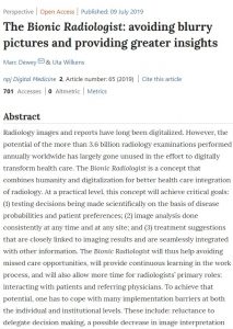
The Bionic Radiologist: avoiding blurry pictures and providing greater insights.
Prof. Dr. Marc Deweys and Uta Wilkens latest realease helps at a practical level to achieve the following critical goals: (1) testing decisions being made scientifically on the basis of disease probabilities and patient preferences; (2) image analysis done consistently at any time and at any site; and (3) treatment suggestions that are closely linked to imaging results and are seamlessly integrated with other information.
Conclusion:
The Bionic Radiologists promises better outcomes and lower costs through better integration of diagnostic imaging in clinical care processes. In this perspective we outlined the characteristics and facilitating factors with respect to the technology development itself, the surrounding institutional conditions and resistance due to the required change in culture as well as the prerequisites for individual acceptance and adaptation.
The Bionic Radiologist will thus help avoiding missed care opportunities. Read more…
July 02, 2019
Kidney Injury after Intravenous versus Intra-arterial Contrast Agent in Patients Suspected of Having Coronary Artery Disease: A Randomized Trial
Eva Schönenberger, Peter Martus, Maria Bosserdt, Elke Zimmermann, Rudolf Tauber, Michael Laule and Prof. Marc Dewey teamed up to compare intravenous versus intra-arterial contrast agent administration in relationship to AKI and analyze the association between AKI and chronic kidney disease.
The results and conclusion:
A total of 320 participants (163 [50.9%] women; mean age, 60 years ± 11) were included. Baseline eGFR did not differ between the CT angiography group (84.3 mL/min/1.73 m2 ± 17.2) and the catheterization group (87.1 mL/min/1.73 m2 ± 16.7) (P = .14). AKI occurred in nine of 161 participants in the CT angiography group (5.6%; 95% CI: 3%, 10%) and in 21 of 159 participants in the catheterization group (13.2%; 95% CI: 9%,19%) (relative risk, 2.4; 95% CI: 1.1, 5.0; P = .02). Also in the subgroup of participants without obstructive CAD, in those not requiring coronary interventions, AKI was more common in the catheterization group (11.9%; 95% CI: 8%, 19%) than in the CT angiography group (4.3% [95% CI: 2%, 9%]; difference, 7.7% [95% CI: 1.3%, 14.1%]; relative risk, 2.8 [95% CI: 1.1, 7.0]; P = .02). Obstructive CAD (odds ratio [OR]: 2.7 [95% CI: 1.1, 6.6]; P = .02), femoral catheter access (OR: 2.5 [95% CI: 1.1, 5.6]; P = .04), and cine ventriculography were associated with AKI (OR: 2.3 [95% CI: 1.0, 4.9]; P = .03). In multivariable analysis, the presence of postcontrast AKI was associated with chronic kidney disease (hazard ratio: 12.4 [95% CI: 4.5, 34.6]; P < .01).
Acute kidney injury was more common after cardiac catheterization than after CT angiography in this prospective randomized study of patients suspected of having coronary artery disease.
Read about this release here.
June 15, 2019
Summer excursion to Prenzlauer Berg
The Dewey AG is not only researching the hottest topics of radiology, but we’re also curious explorers of our city and our neighbourhood of Prenzlauer Berg. The home district of Prof. Dewey was part of a guided summer excursion with many interesting and little known facts about this very historical part of Berlin.
It was a great saturday, we are looking forward to the next excursion!
[ngg src=”galleries” ids=”3″ display=”basic_thumbnail” ajax_pagination=”0″ show_all_in_lightbox=”0″ use_imagebrowser_effect=”0″ display_view=”default”]
June 12, 2019
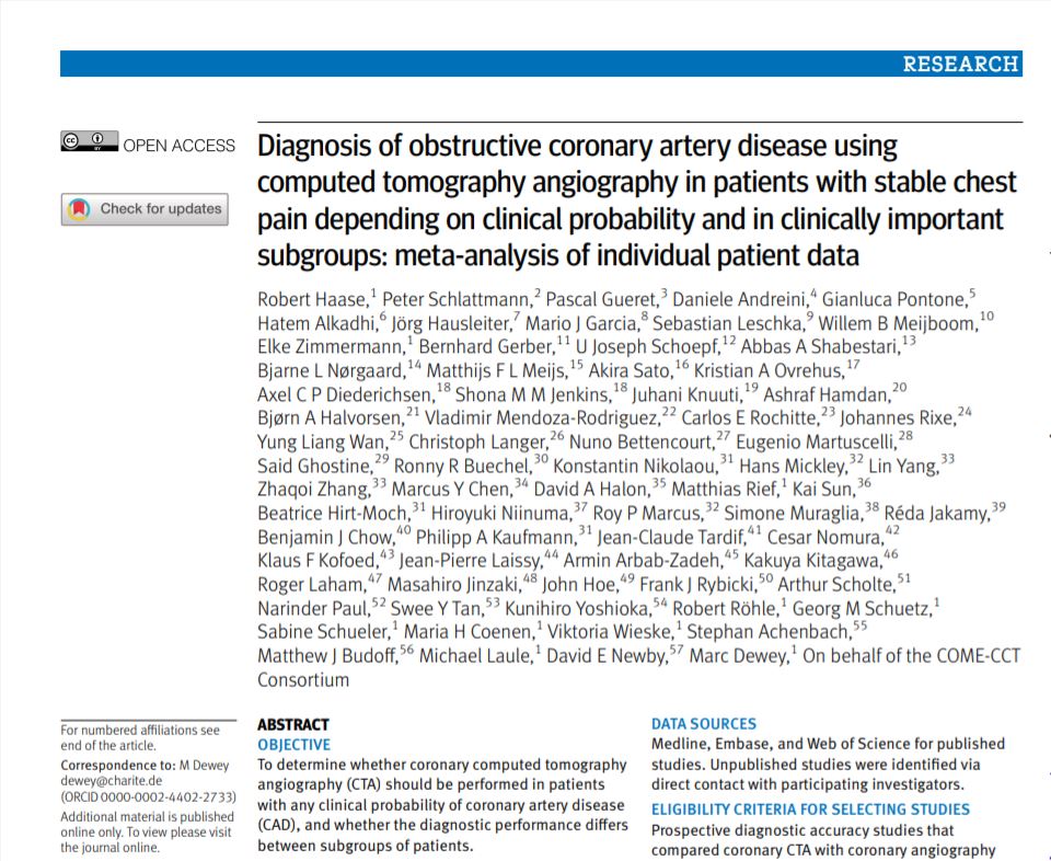
The latest scientific release from Mr. Robert Haase by the british Medical Journal on the topic of “Diagnosis of obstructive coronary artery disease using computed tomography angiography in patients with stable chest pain depending on clinical probability and in clinically important subgroups: meta-analysis of individual patient data”.
The objective was to determine whether coronary computed tomography angiography (CTA) should be performed in patients with any clinical probability of coronary artery disease (CAD), and whether the diagnostic performance differs between subgroups of patients. Read more about it here.
May 22, 2019
Prof. Dr. Udo Hoffmann, Professor of Radiology at Harvard Medical School and Chief of Cardiovascular Imaging at Massachusetts General Hospital (Boston, USA) has been selected in a new round of BIH visiting professor approvals. Together with the two hosts from the Radiology Department of the Charité, Professor Dr. Bernd Hamm and Professor Dr. Marc Dewey, their mutual aim is the preparation of an international research network for the joint use of imaging data (GUIDE-IT). We’d like to say congratulations and we’re eager to work with you!
About Prof. Dr. Hoffmann:
Udo is a pioneer and world leader in cardiovascular imaging. He leads a vibrant multidisciplinary research program at MGH focused on translational and clinical research to utilize cardiovascular imaging to improve prevention, diagnosis, and treatment of cardiovascular disease. Dr. Hoffmann has led hallmark randomized clinical trials in CV imaging, including ROMICAT I and II, PROMISE, and REPRIEVE. He published more than 500 peer reviewed manuscripts is among the top 1% cited researchers in his field. He received the SCCT Gold Medal in 2018.
Read more about the new funding rounds at the Informationsdienst der Wissenschaft here.
May 22, 2019
https://youtu.be/33hpYvYsDu8
The Dewey AG Team was involved in the Discharge trial, coordinated by Prof. Dr. Marc Dewey. The Discharge trial is a collaborative multinational research project project which examines for which patients with suspected coronary artery disease based on stable chest pain, cardiac computed tomography (CT) or cardiac catheterisation is best suited. (Video in German language.)
May 21, 2019
[kad_youtube url=”https://www.youtube.com/embed/XwgwD1dq3Fo” ]
A short teasing video from last years Pre-Research Forum with Prof. Dr. Marc Dewey being interviewed and asked about the upcoming questions of new health and artificial intelligence becoming a big part of radiology in the future.
May 9, 2019
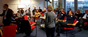
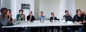
The AG Dewey is officially back!
We are back in Berlin Mitte, inspired to drive things forward in todays medicine and radiology. The two days in the retreat held many informative speeches for the whole group and we are especially thankful to your guestspeakers who joined us in the last days. We would like to say thank you to Prof. Gabriel Krestin, Prof. Göran Bergström and Prof. Sebastian Stober, who were talking exciting topics such as artificial intelligence, shared their experiences with us and generally motivated enormously. And of course thanks to our Prof., Mr. Marc Dewey for making this event happen!
Take a brief look at the speeches of the team Dewey and our great guests:
[ngg src=”galleries” ids=”1″ display=”basic_imagebrowser”]
April 27, 2019
The AG Dewey is officially retreating!
But only for a short time, to have some informative and inspiring days with

lectures, presentations and discussions from speakers of the AG Dewey as well as several guests. From Tuesday, 07.05.2019 until Wednesday, 08.05.2019 we will be residing in the Seminaris Hotel near Potsdam and are eager to hear about the most current and driving topics of radiology and the AG Dewey!
A rough overview about the topics of the presentations:
- Clinical Trials
- Core320
- CAD-Man
- Discharge
- ComeCCT
- Artificial Intelligence
- Claustrophobia
- Perspectives
- Promotionskolleg
March 26, 2019
The following feels like a great compliment to all of our team, since we are always very eager to drive forward scientific research, medicine and technology here at the radiology department of the Charité in Berlin Mitte. The Charité has been mentioned within newsweek.com’s TOP 10 best hospitals in the world and Charité even makes it into their Top 5! Together with Statista Inc, a global market research and consumer data company, newsweek.com developed a groundbreaking ranking of the world’s best hospitals. Hospitals on this list are at the forefront of adapting to new challenges while providing top-notch patient care.
About the Charité Berlin newsweek.com states: “As a hospital, Charité—which celebrated its tricentennial in 2010—is on the cutting-edge of biomedical innovation, with biotech startup labs, advisory roles and business initiatives focused on the convergence of technology and medicine.”
Thank you for this compliment, we will keep giving our very best. Read the whole article by Noah Miller …
March 24, 2019
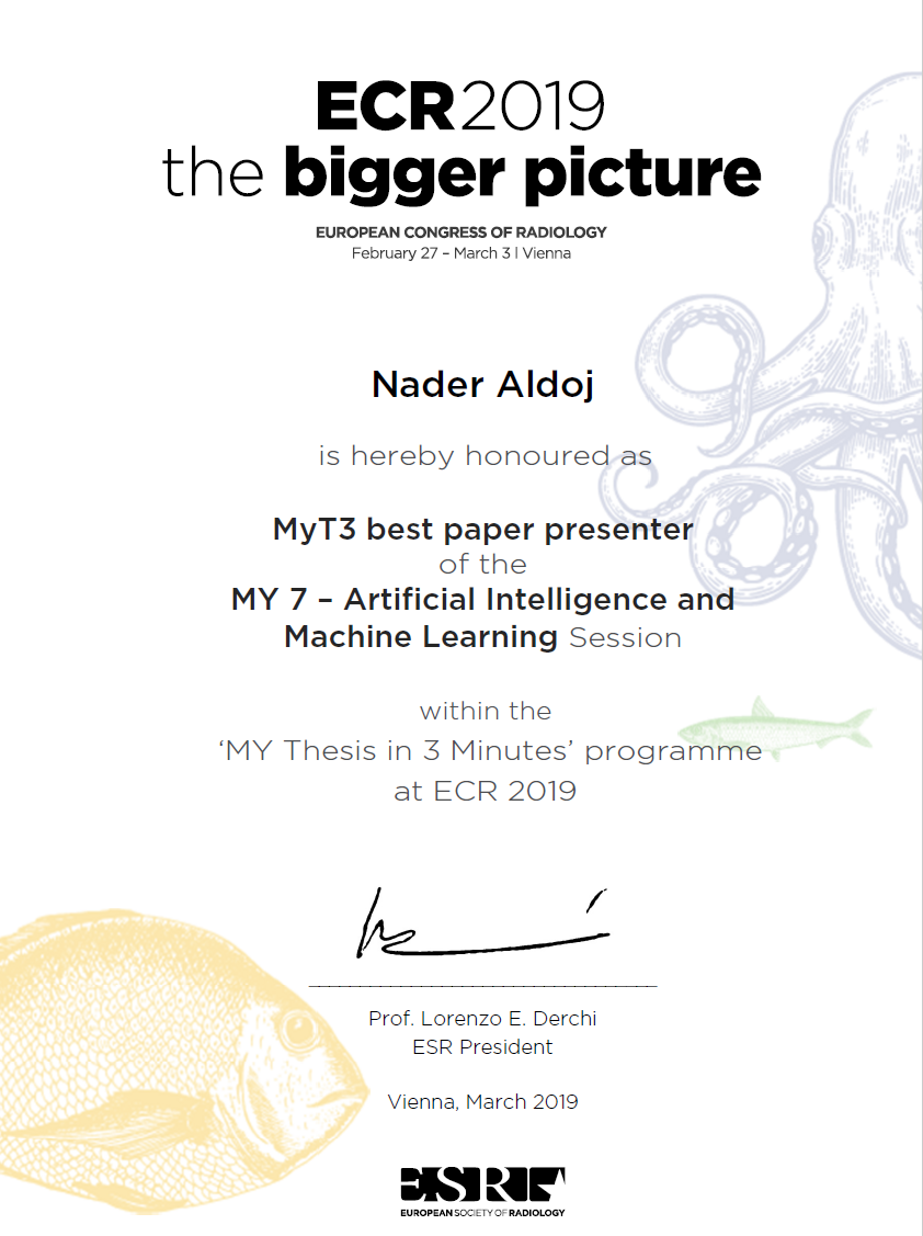
Prof. Dewey and the other group members congratulate Nader Aldoj on being awarded the best paper presentation of the “My Thesis in 3 Minutes” Session MY 7 at the ECR 2019.
March 22, 2019
A current publication under participation of the radiology department of Charité under Prof. Dr. Marc Dewey takes on “Patient Preferences for Coronary CT Angiography with Stress Perfusion, SPECT, or Invasive Coronary Angiography”.
In a multicenter prospective study, patients had a two- to threefold preference for coronary CT angiography with stress perfusion over SPECT and invasive coronary angiography for evaluation of coronary artery disease.
Read more about it here.
Also take a look at the paper about “preferences in cardiac testing” and the question “Why patient experience matters” from the Mallinckrodt Institute of Radiology by Pamela K. Woordward and Thuy D. Nguyen in this publication.
March 15, 2019
Interview of Prof. Dr. Marc Dewey by Michael Reiter on radiological studies at Universitätsmedizin Charité Berlin. Learn about the DISCHARGE study and much more about what is going on in the Charité radiology department in the video.
March 8, 2019
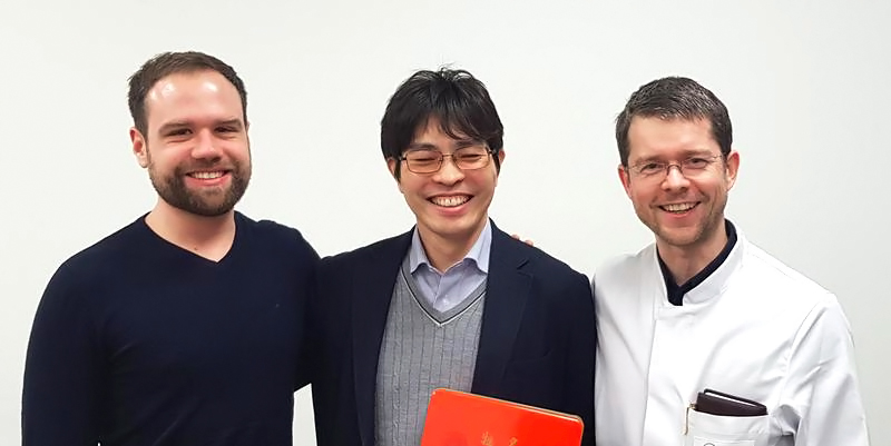
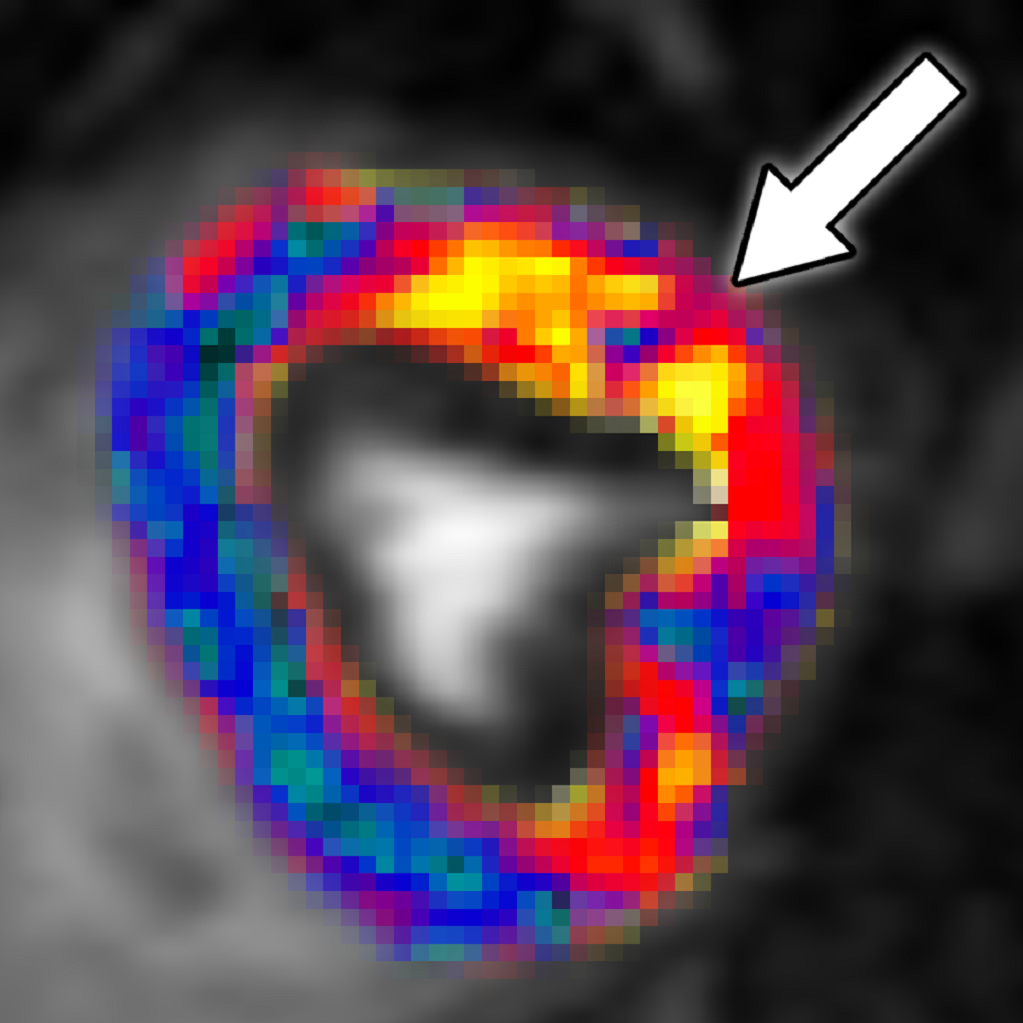
The project “Fractal Analysis of Myocardial Ischemia” of the Deutsche Forschungsgemeinschaft (DFG, German Research Foundation) applied successfully for a Mercator-Fellow – the first ever in Radiology. Kakuya Kitagawa, MD, PhD, of the Department of Radiology at the Mie University Hospital in Japan will be supporting the project with his expertise and his international perspective.
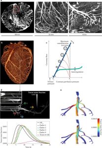
The DFG-project “Fractal Analysis” was proposed by Prof. Dr. Marc Dewey and Dr. Florian Michallek from the department of Radiology at Charité Universitätsmedizin Berlin. The researchers want to develop a method to examine the causes of chronic ischaemic heart disease in a better and non-invasive way. They investigate if fractal analysis is a comprehensive perfusion analysis method (Figure 1). “We are looking forward to the scientific contribution of Kakuya to this project as he is one of the most renown experts on multimodality myocardial perfusion imaging worldwide.” says Prof. Dewey and Dr. Michallek explains “The Mercator-Fellowship Programme allows on the one hand to involve the distinguished expertise of Prof. Kitagawa for the project and on the other hand to synthesise a highly interesting and multi-centric dataset.
(From IDW-online press release)
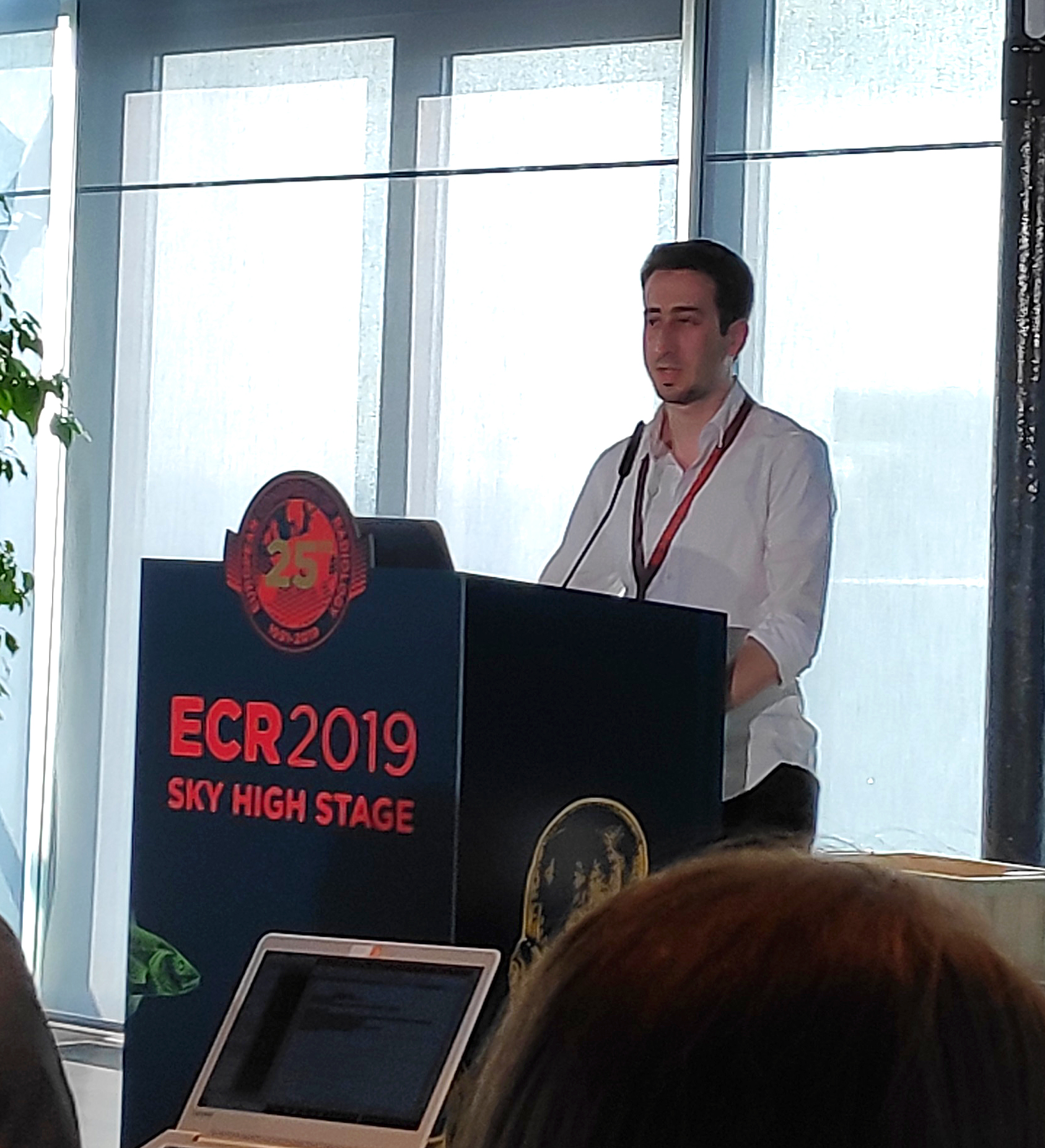
February 27, 2019
The European Congress of Radiology (ECR) 2019 has just started in Vienna on February 27, 2019. We have seen great presentations in the sky-high stage (see picture), among them the publication by Dr. Bosserdt in Eur Radiol (link) about the ESR survey on imaging trials and data sharing. The group also enjoyed the small party at Henglbrunner (see picture).

January 30, 2019
Publication of “Impact and perceived value of journal reporting guidelines among Radiology authors and reviewers” in European Radiology by Marc Dewey, Deborah Levine, Herbert Y. Kressel and Marc Bossuyt.
September 28, 2018
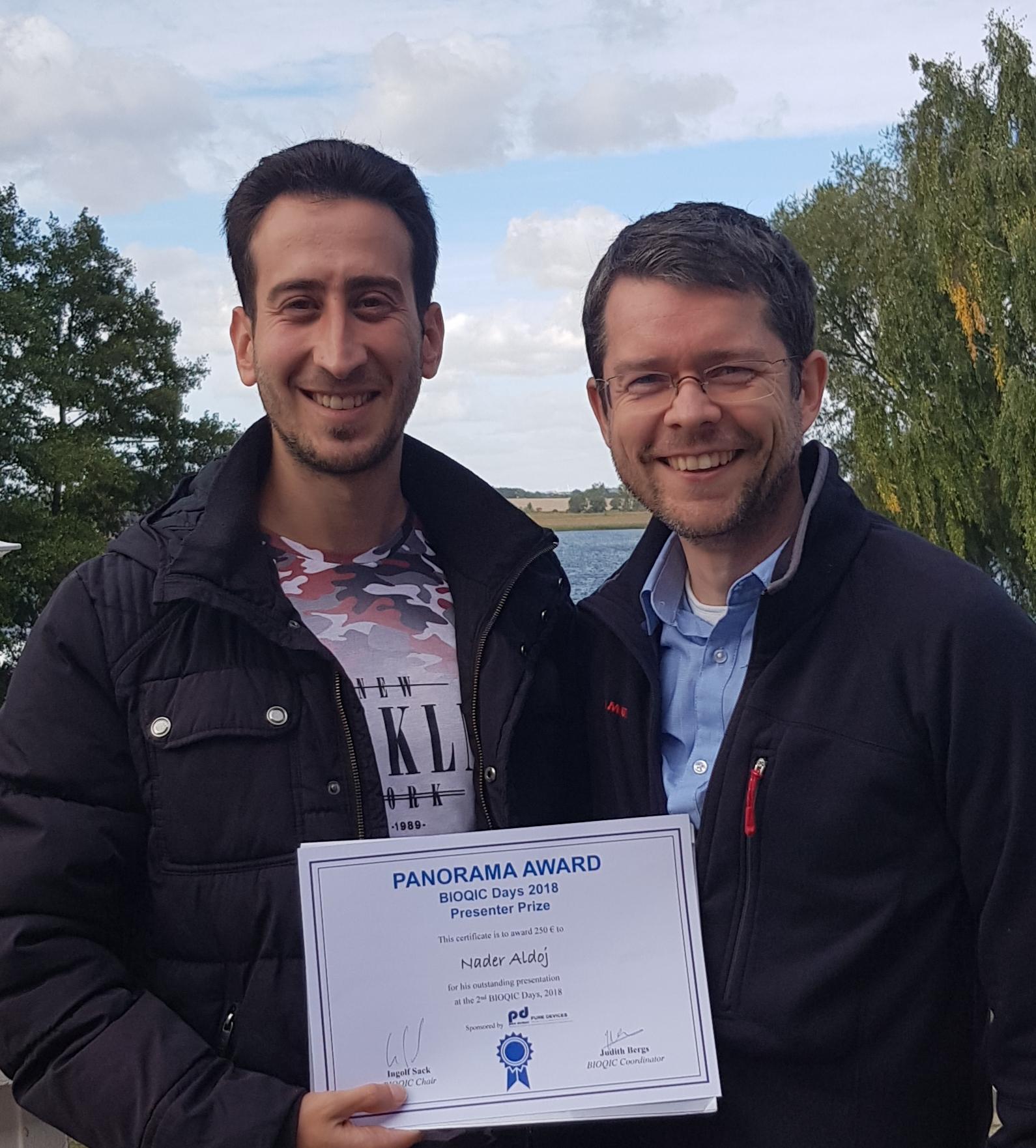
This years BIOQIC best presentation award went to Nader Aldoj. Congratulations! .