
Research Projects
Noninvasive imaging has benefited from technical advances that might allow it to play a pivotal role in cardiovascular diagnosis and patient management. Most notably, advanced imaging modalities have great potential for noninvasive characterization of coronary atherosclerosis as well as quantification of myocardial perfusion.
Motivating national and international collaboration is one of the central aims of our working group. We have initiated two international multicontinental consortia that perform exciting research on cardiovascular imaging in patients with suspected coronary artery disease (COME-CCT) and patients who already have coronary stents (COME-CSI).
Furthermore, Prof. Marc Dewey’s working group has received an EU Framework grant for the DISCHARGE project. This group of 30 academic partners from 18 countries is coordinated by Marc Dewey and allows the investigators to raise awareness among patients, health care providers, and decision-makers about the effectiveness and cost-effectiveness of coronary CT angiography. Main results of the trial were published in NEJM and BMJ.
Recently, Prof. Dewey`s working group received funding by the German Research Foundation (DFG) for the project Guide to Data Sharing of Imaging Trials (GUIDE-IT). Aim of the 2-year pre-project which started in May 2023 is to develop a concept for an infrastructure for sharing data from randomized medical imaging trials. The work packages leaders and/or collaborators in the project are Dr. Melanie Estrella and Dr. Maria Bosserdt (both from our group) and Prof. Felix Balzer (Charité), Prof. Dagmar Krefting (UMG Göttingen) and Prof. Martin Gersch (FH Berlin). In a subsequent full-project phase, the best-suited concept will be implemented provided funding will be granted.
Below you can find a selection of our recent and current research projects:
2023-2025 GUIDE-IT Project
Randomized imaging trials compare clinical outcomes of two or more medical imaging strategies and are thus crucial for advancing medical care and guidelines in this field. To facilitate data sharing for these types of studies, large and challenging data sets have to be curated. In the GUIDE-IT project concepts to overcome these barriers will be developed, also including stakeholder acceptance, use and access, data quality, IT resources and economic sustainability. The aim is to create an infrastructure, which is tailored to the needs of the community of data providers and re-users, as well as additional stakeholders as patients and policy makers. Enabling data sharing for this type of data would also advance the development of novel imaging biomarkers using artificial intelligence and radiomics.
Read more: https://marcdewey.de/successful-guide-it-kick-off
2020 – 2023 Radiomics: Next Generation of Biomedical Imaging
Priority Programme Radiomics for the investigation of coronary flow.
Background and Objectives: Coronary artery plaques may lead to cardiovascular events and there is early evidence that noninvasive detection of vulnerable plaque features on computed tomography (CT) and subsequent changes in medical management may lead to improved outcomes. CT is an increasingly used test in patients with suspected coronary artery disease (CAD) and is the best noninvasive imaging test to capture three-dimensional information about plaques in all coronary artery segments in one examination. The quantitative results of image analysis will be made available as a database with cardiovascular events.Anticipated Gain of Knowledge: We anticipate novel insights into the association of coronary plaque features with prognosis of stable chest pain patients which will strengthen the clinical implications of radiomics analysis and deep learning of coronary CT. Ultimately, this will allow identifying patients prone to suffer myocardial infarction and testing the validity and generalisability of advanced coronary CT plaque analysis using data from the population-based SCAPIS project of more than 25000 asymptomatic individuals which will have long-term follow-up data for the second three-year funding period.
SPP (Priority Program) Radiomics Plaque: https://gepris.dfg.de/gepris/projekt/428222922
SPP Radiomics Flow: https://gepris.dfg.de/gepris/projekt/428223139
Since 2018 Fractal Analysis Project
The DFG-project “Fractal Analysis” driven by Prof. Dr. Marc Dewey and Dr. Florian Michallek as well as the Mercator Fellowship Professor Kakuya Kitagawa.

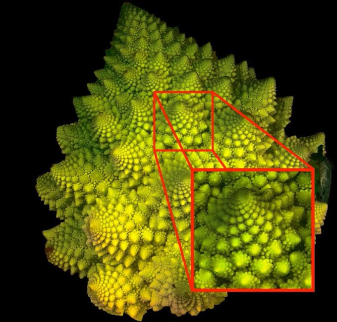
The project “Fractal Analysis of Myocardial Ischemia” of the Deutsche Forschungsgemeinschaft (DFG, German Research Foundation) applied successfully for a Mercator-Fellow – the first ever in Radiology. Kakuya Kitagawa, MD, PhD, of the Department of Radiology at the Mie University Hospital in Japan will be supporting the project with his expertise and his international perspective. The DFG-project “Fractal Analysis” was proposed by Prof. Dr. Marc Dewey and Dr. Florian Michallek from the department of Radiology at Charité. The researchers want to develop a method to examine the causes of chronic ischaemic heart disease in a better and non-invasive way. They investigate if fractal analysis is a comprehensive perfusion analysis method (Figure 1). “We are looking forward to the scientific contribution of Kakuya to this project as he is one of the most renown experts on multimodality myocardial perfusion imaging worldwide.” says Prof. Dewey and Dr. Michallek explains “The Mercator-Fellowship Programme allows on the one hand to involve the distinguished expertise of Prof. Kitagawa for the project and on the other hand to synthesise a highly interesting and multi-centric dataset.”
More information: https://gepris.dfg.de/gepris/projekt/392304398
2017-2021 BIOCIQ – BIOphysical Quantitative Imaging Towards Clinical Diagnosis

The BIOQIC research program will advance medical imaging methods towards the system-independent quantification of the constitution and structure of soft tissues. In-depth training will be provided through block courses in ‘Imaging Physics & Chemistry’, ‘Biophysics & Biomechanics’ and ‘Mathematics & Signal Processing’. The program is specifically designed to provide training in imaging sciences and to enhance skills for becoming an independent researcher.
Grants: https://gepris.dfg.de/gepris/projekt/289347353
Grants: https://gepris.dfg.de/gepris/projekt/428223139
Since 2017 – 2018 BIH Digital Health Innovator Program
For Digital Health Innovators at BIH, Charité and MDC.
The BIH Digital Health Accelerator Program supports all employees from research and clinical departments of the Charité including the BIH in developing digital health solutions from their innovative ideas and transferring them to patients in medical applications or to the healthcare market.
Our working definition of Digital Health is the convergence of digital technologies such as 3D printing, augmented/mixed/virtual reality, machine learning/artificial intelligence, robotics, sensors and software with life sciences and healthcare. While the boundary with medical technology, diagnostics, etc. is sometimes blurred; key is the presence of digital technology and data use as core components of the innovation.
Participating project teams advance through the Digital Health Accelerator program along a structured process for iterative product development supported by:
- Funding (including Protected Time for a fraction of time off from the clinic).
- Mentoring by high-profile national and international experts Access to networks (talents, development partners, industry, investors)
- Interdisciplinary BIH DHA team BIH Digital Labs co-working space near the Charité Campus Mitte.
2014-2020 EU DISCHARGE project
DISCHARGE is a collaborative multinational research project. The consortium of the project is composed of 31 members in 18 European states. Core of the project is a pragmatic randomised controlled trial which includes 26 clinical sites from 16 European countries. The project examines for which patients with suspected coronary artery disease based on stable chest pain, cardiac computed tomography (CT) or cardiac catheterisation is best suited and is based on the single-centre experience with the CAD-Man study at Charité coordinated by Prof Dewey.
DISCHARGE has the capability to influence current standards and guidelines as well as coverage decisions and will raise awareness among patients, health care providers, and decision-makers in Europe about the effectiveness and cost-effectiveness of cardiac CT. The project was funded by the European Commission and is coordinated by the Department of Radiology of Charité – Universitätsmedizin Berlin (coordinator: Prof. Dr. Marc Dewey).
The DISCHARGE trial has published its main analyses in NEJM and BMJ. We accept applications from MD and PhD doctoral students for positions in our group, please do not hesitate to make contact with your CV and motivation using dewey@charite.de.

The DISCHARGE Trial results on gender differences were published in The British Medical Journal (The BMJ). Thanks to all members of the team, and of course to the patients participating in the trial.

2013-2015 BMBF/DFG
“CoMeCSI Consortium- Collaborative Meta-Analysis of Coronary Stent Imaging“

2012-2013 BMBF/DFG

Clinical Assessment of Coronary Computed Tomography Angiography: Collaborative Meta-Analysis of Individual Patient Data from Prospective Diagnostic Accuracy Studies“
2011-2012 DFG
“Prediction of Claustrophobia during MR imaging”

2010-2011 BMBF/DFG
“Meta-Analysis of noninvasive coronary angiography using multislice computed tomography for detection of coronary in-stent restenosis”.

2011-2012 German Heart Foundation and German Foundation for Heart Research:
“Evaluation of coronary atherosclerosis using 320-row CT and comparison with urine proteome analysis“
2009-2020 Toshiba Medical Systems:
„CORE-320“

2010-2012 Hypatia Program:

“Reduction of Claustrophobia during MR imaging”
2009-2010 German Heart Foundation and German Foundation for Heart Research:

„Noninvasive coronary angiography using 320-row CT“
2009-2012 EFRE:
„Software based noninvasive analysis of cardiac function using multislice computed tomography
